
Figure 5. HCT-15 cells have higher basal level of topoI-pS10 and generating topoI-EGFP fusion cells.
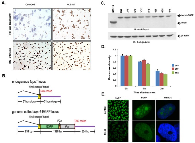
A. Colo 205 and HCT-15 cell pellets were fixed and subsequently embedded in paraffin. Slides were cut from the cell pellet and IHC staining with anti-topoI-pS10 (upper panel) and anti-topoI (lower panel) was performed. B. A SpCas9-VQR variant plasmid and a sgRNA expression plasmid targeted to the hTOP1stop codon were co-transfected with a homologous recombination donor plasmid in HCT-15 cells to generate topoI-EGFP fusion protein expressing cell lines. C. Single EGFP positive cells were sorted and grown, and several clones were selected and characterized by western-blot. Cells lysates were subjected to immunoblot analysis with anti-topoI (upper panel) and anti-β-actin (lower panel). D. Three clones (3H8, 4E7 and 4H8) were further characterized by determining topoI degradation. Cells were treated with 2.5μM SN38 for 3h and EGFP florescence intensity was quantitatively analyzed by plate reader. E. Cells from clone 4E7 (HCT-15 4E7) were treated with 2.5μM SN38 (active metabolite of irinotecan) for 3h. Cells were visualized by using a Leica S5 confocal microscope.