
Figure 2. Affinity purification of TAT-BMI-1 and TAT-GFP recombinant proteins.
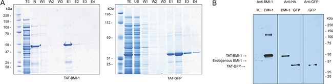
(A) Coomassie staining of NUPAGE 4–12% gels of total induced bacterial extract (TE), input (IN), unbound (UB) wash (W) and Imidazole elution (E). (B) Western Blotting of purified TAT-proteins, identified by specific antibodies against the HA epitope or for BMI-1 and GFP. A total extract from K562 shows the presence of native BMI-1 at 36.9 KDa compared to the TAT-BMI-1 at 45 KDa.