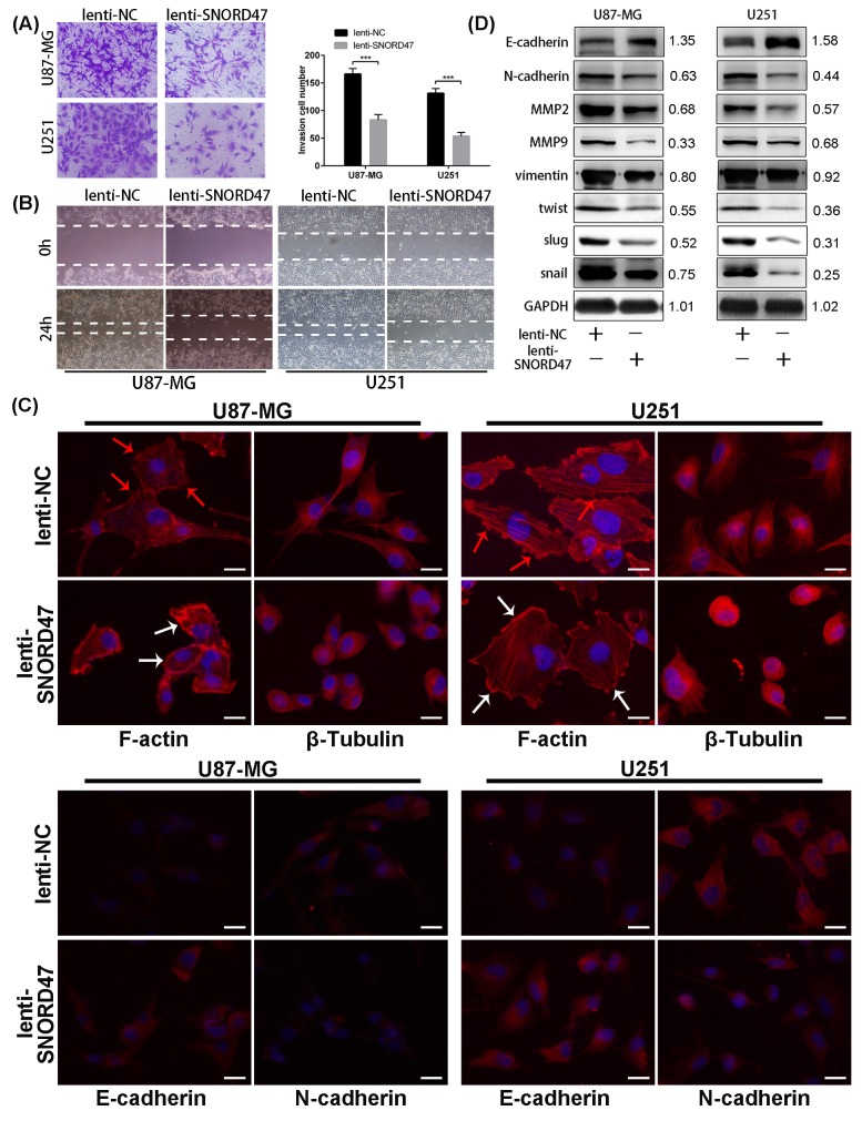
Figure 4. SNORD47 suppressed the invasion and epithelial-to-mesenchymal in glioma cells.
(A) Invasion ability of glioma cells with or without SNORD47 overexpression glioma cells was measured by transwell assay. ***P<0.001 (Student's t test). (B) The cell migration of U87-MG and U251 cell line was examined by wound healing assay. (C) The results of F-actin staining displayed a cortical pattern in lenti-SNORD47-treated cells and a stress-fiber pattern in Lenti-NC-treated cells. β-Tubulin staining showed a skeleton retraction after treated with SNORD47. Immunofluorescence staining also displayed a lower N-cadherin expression level and a higher E-cadherin expression level after the treatment with lenti-SNORD47. (D) Western blot analysis exhibited the expression of EMT-associated proteins in lenti-NC or lenti-SNORD47 treated glioma cells. The red arrow and the white arrow indicatedinvadopodia and lamellipodia respectively. Bar =20μm.

