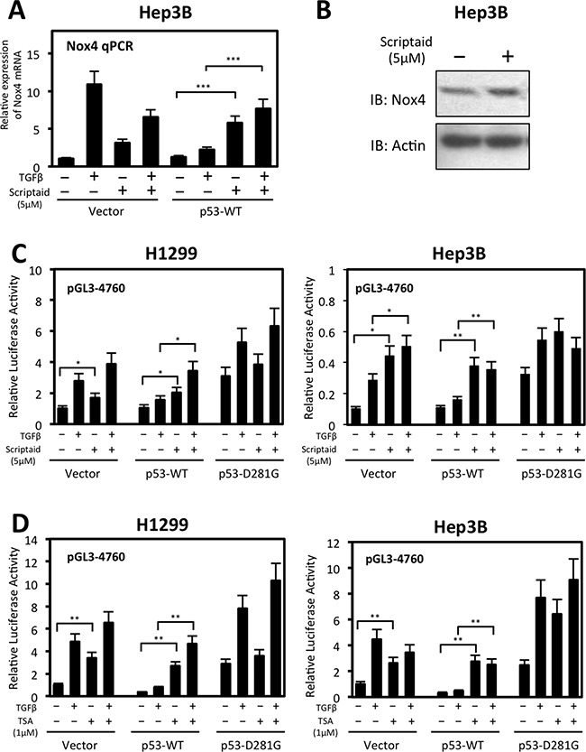
Figure 8. Histone deacetylase (HDAC) activity participates in wild-type p53-mediated repression of NOX4 mRNA and promoter activity.

(A) Hep3B cells were transiently transfected with control vector or p53-WT. Twenty-four hours after transfection, cells were treated with TGFβ (5 ng/ml) and either Scriptaid (5 μM) or DMSO for 24 hours. Real-time qPCR analysis of NOX4 mRNA expression was detected with NOX4-specific primers. The relative mRNA level was normalized to GAPDH control. (n = 4) (B) Hep3B cells transfected with empty vector were treated with Scriptaid (5 μM) for 24 hours. After 24 hours, total cell lysates were collected and analyzed by immunoblot. (C) H1299 or Hep3B cells were co-transfected with pGL3-NOX4 (-4760) and either vector control, p53-WT, or p53-D281G. Twenty-four hours later, cells were left untreated or treated with TGFβ (5 ng/ml) and either Scriptaid (5 μM) or DMSO (-) for another 24 hours. Total cell lysates were then collected and assayed for luciferase activity. (D) Luciferase assays were conducted on H1299 or Hep3B cells co-transfected with pGL3-NOX4 (-4760) p53 plasmids as in B. Twenty-four hours later, cells were left untreated or treated with TGFβ (5 ng/ml) and either trichostatin A (TSA) (1 μM) or DMSO (-) for 24 hours (n = 3, in triplicate). Significance values are indicated as *P-value < 0.05, **P-value < 0.01, or ***P-value < 0.001.
