
Figure 4. IL-17 reduces oxaliplatin-induced apoptosis in HCC cells.
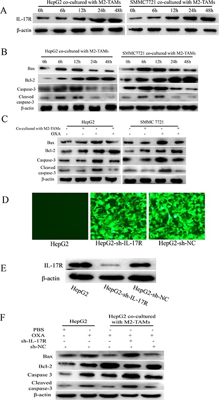
(A and B) HepG2 and SMMC-7721 cells were co-cultured with M2-TAMs for 24 h and then treated with 20 μg/mL oxaliplatin for various times. IL-17R (A), BAX, BCL-2, caspase-3 and cleaved caspase-3 (B) were detected by Western blotting. (C) HepG2 and SMMC-7721 cells were cultured with or without M2-TAMs for 24 h and then treated with 20 μg/mL oxaliplatin for another 24 h. BAX, BCL-2, caspase-3 and cleaved caspase-3 were detected by Western blotting. (D and E) HepG2 cells were infected with lentiviruses expressing specific shRNAs (sh-IL-17R or sh-NC). The transfection efficiency was detected by fluorescence microscopy (200×) (D) and the knockdown efficiency was detected by Western blotting (E). (F) sh-IL-17R HepG2 cells were cultured with or without M2-TAMs for 24 h and then treated with 20 μg/mL oxaliplatin for another 24 h. BAX, BCL-2, caspase-3 and cleaved caspase-3 were detected by Western blotting.