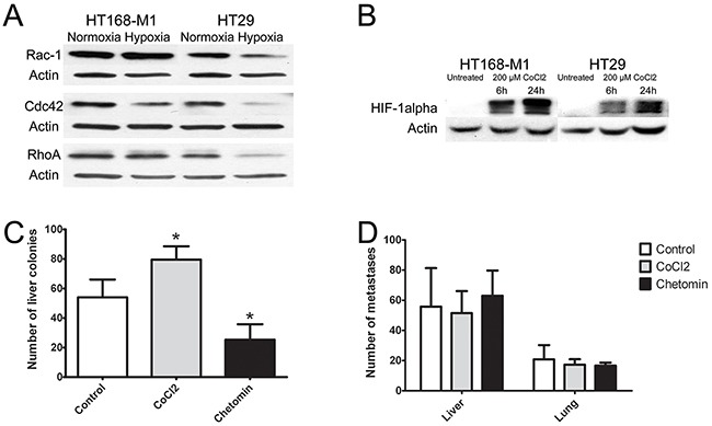
Figure 3. Effect of hypoxia on protein expression and in vivo metastasis of human tumor xenografts.

(A-B) Protein expression of small GTPases and HIF-1α in HT168-M1 and HT29 cells under hypoxic conditions (1% O2 level) compared to normoxia (a representative blot). (C) HT168-M1 human melanoma cells were injected intrasplenically and liver colonies formed were counted at day 34 of the treatment. (D) HT29 human colon cancer fragments were orthotopically transplanted to the appendix region and liver and lung metastases were counted at day 34. Note that the CoCl2 treatment increased metastasis formation by the highly motile HT168-M1 cells. Inhibition of HIF proteins by chetomin, on the other hand, resulted in a significant decrease in the metastatic potential. However, CoCl2 or chetomin treatment had no effect on the metastatic capacity of HT29 cells. Data represent mean ± SEM of two independent experiments; *p<0.05.
