
Figure 3. BRCA2 moves to an insoluble fraction after treatment with MG132 or 44 °C.
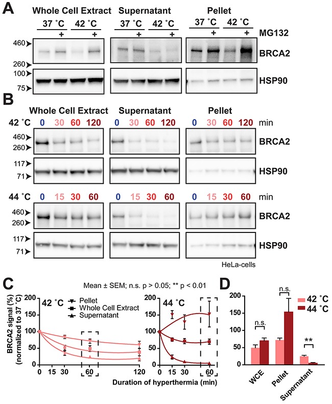
(A) BRCA2 protein levels at 37 °C and 42 °C in HeLa cells treated with or without 50 μM MG132 on cropped immunoblots. Signals shown are from the whole cell extract (left), and from two fractions after centrifugation: the supernatant (middle) and in the pellet fraction (right). HSP90 is used as a loading control. (B) BRCA2 protein levels on cropped immunoblots at 42 °C (upper panel) and 44 °C (lower panel) in HeLa cells in the whole cell extract (left), and from the two fractions after centrifugation: the supernatant (middle) and in the pellet fraction (right). HSP90 is shown as a loading control. (C) Quantification of BRCA2 signals in B, corrected for the 37 °C control. A linear-quadratic regression indicates three separate curves for each series of measurements, indicating a significant difference in BRCA2-protein levels in the whole cell extract, supernatant and pellet. The error bars denote mean ± SEM obtained in three experiments. (D) Bars zoom in on the 60-minute treatment point in C. Statistical differences of column pairs were determined using a student's t-test.