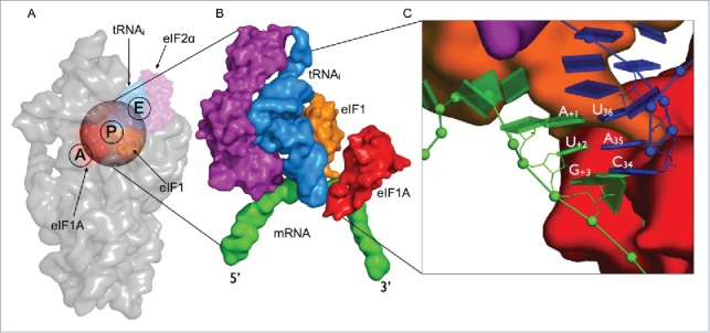
Figure 1.

Structural overview of the eukaryotic initiation complex (A) Small subunit of the eukaryotic ribosome (gray) showing the initiation factors eIF1 (red), eIF1A (orange) and eIF2-α (purple) in complex with initiator tRNA (blue) bound to the AUG start codon (green). The transparent sphere on top of the P-site is the volume where the simulations are focused. (B) A close-up view onto the mRNA-tRNAi complex in surface representation illustrating the proximity of the initiation factors to the codon-anticodon minihelix. (C) A zoomed in view of the codon-anticodon triplet is represented as blocks using the standard reference frame for nucleic acids. eIF1 can be seen to bind closer to the first codon position toward the E-site while eIF1A is located in the A-site closer to the third codon position. The rRNA and proteins are omitted for clarity.
