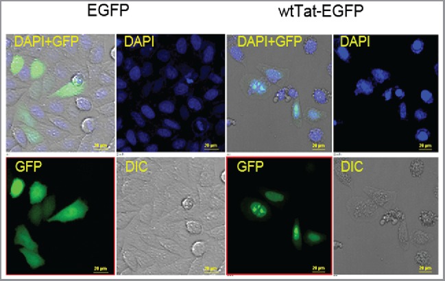
Figure 1.

Subcellular localization of Tat-EGFP fusion protein folding in HeLa cells. HeLa cells were transfected with EGFP or wild type (WT) EGFP-Tat fusion constructs, and subcellular localization was determined by imaging EGFP using a fluorescence microscope (40X). The EGFP is present in the cytosol as evident in the merged (DAPI + GFP) image. Tat-EGFP localizes predominantly in the nucleus, frequently as nuclear speckles. GFP filtered images correspond to the location of EGFP or Tat-EGFP protein expression. DAPI stains the nucleus of all fixed cells. The merged image contains the combined images of GFP, DIC (Differential Interference Contrast), and DAPI staining, and shows a representative area under microscopic observation. Scale bar, 20 μm.
