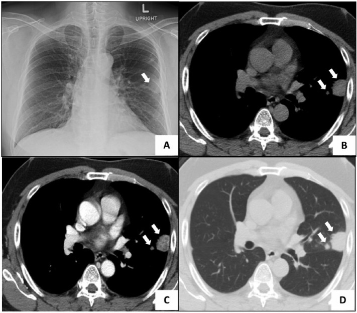
Figure 1.
(A) Chest X-ray revealing a nodule in the middle part of the upper left lung. (B) Mediastinal window nonenhanced CT image showing 2 masses attached to the chest wall (arrow). (C) Mediastinal window enhanced CT image showing heterogeneous enhancement in the nodules (arrow). (D) CT scan of lung window showing 2 masses at the lingular segment of upper left lobe (arrow). CT indicates computed tomography.

