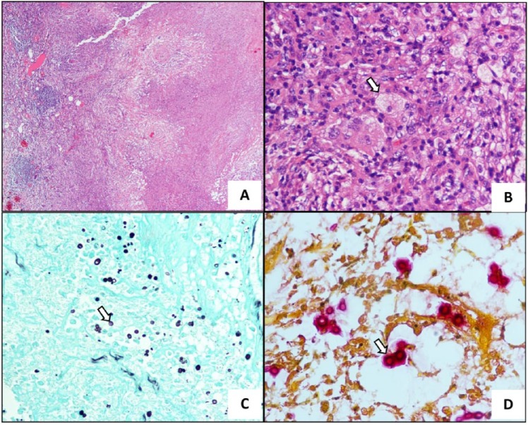
Figure 2.
(A) Histologic slide showing chronic granulomatous inflammation with tissue necrosis (hematoxylin-eosin, original magnification ×40). (B) There were numerous intracellular round-shaped microorganisms in macrophages (arrow) (hematoxylin-eosin, original magnification ×100). (C) Grocott methenamine silver staining demonstrating many yeast-form fungal organisms in the lesion (arrow) (original magnification x100). (D) Mucin staining depicting red-pink color in the capsule of this fungal organism (arrow) (original magnification x400).

