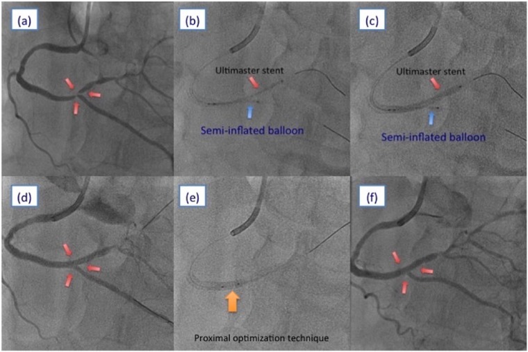
Figure 1.
(a) Right coronary angiogram of the left anterior oblique cranial view. (b, c) Stent implantation using the jailed semi-inflated balloon technique. (d) Right coronary angiogram of the left anterior oblique cranial view after stent implantation. (e) Proximal optimization technique using a non-compliant balloon. (f) Final coronary angiogram.

