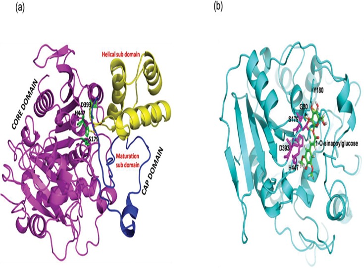
Fig 2. Three dimensional structure and binding of 1-O-sinapoylglucose with SCT enzyme.
(a) Three dimensional structure of SCT enzyme. The core domain is shown in magenta, helical sub-domain in yellow and the maturation sub-domain is highlighted in dark blue. The catalytic triad residues are shown in green sticks. (b) The binding mode of 1-O-sinapoylglucose in deep cavity of SCT enzyme. The substrate 1-O-sinapoylglucose is represented by green stick whereas the cavity of the SCT is shown in cyan colour cartoon. The catalytic triad residues are shown as red sticks. The substrate molecule is hydrogen bonded (yellow dotted lines) with S179, Y180 and G80.

