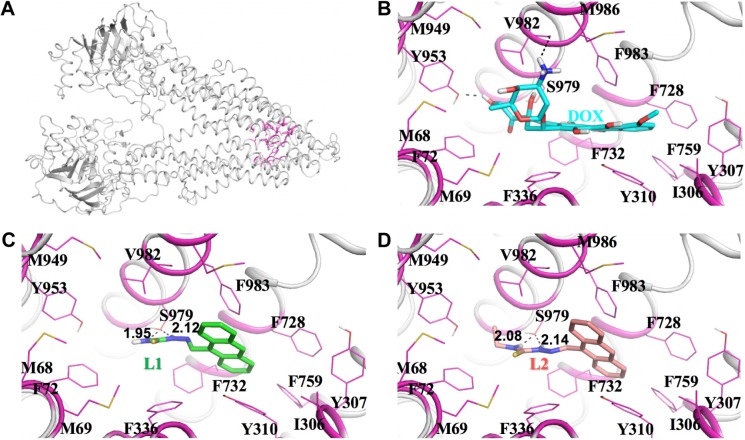
Figure 8.
Predicted binding modes of human P-gp with DOX, L1, and L2.
Notes: The predicted structure of human P-gp (A). The detailed binding interactions of human P-gp with DOX, L1, and L2 (B, C, and D, respectively). Human P-gp is represented as cartoon in gray color. DOX, L1, and L2 are shown in stick. The residues interacting with these molecules are shown in magenta.
Abbreviations: P-gp, P-glycoprotein; DOX, doxorubicin.

