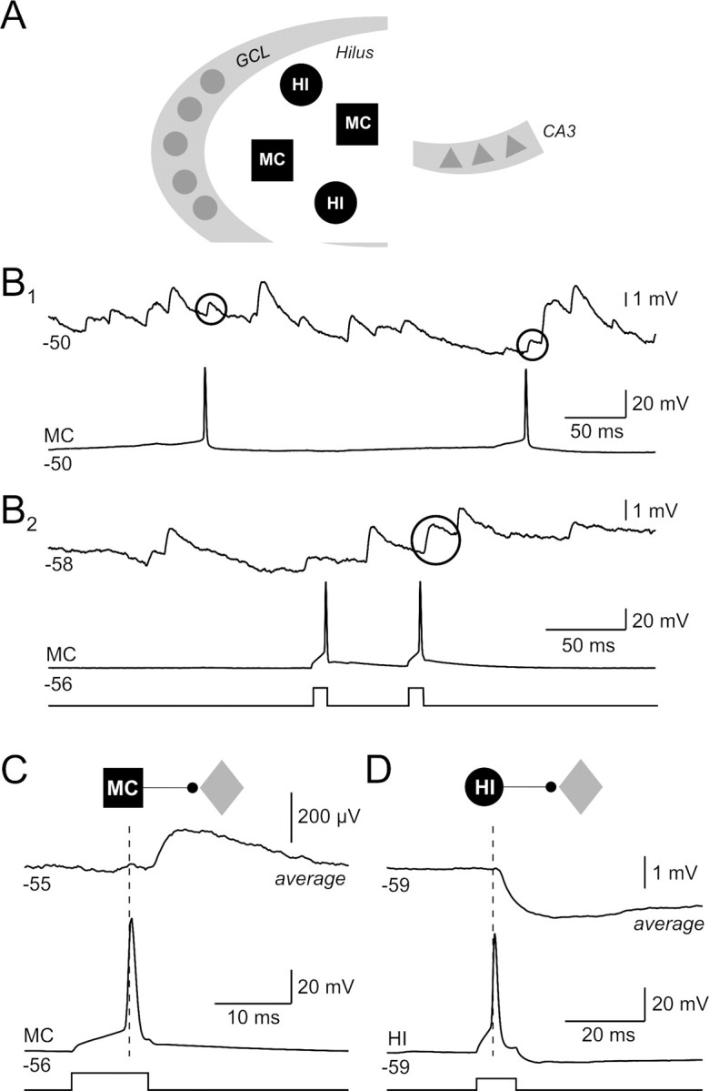
Figure 1.

Synaptic connections formed by dentate hilar neurons. A, Schematic representation of the dentate gyrus and CA3 subfield of the hippocampus. The dentate hilus is located between the granule cell layer (GCL) and CA3 and contains primarily two classes of neurons: MCs and HIs. B1, Paired recording between two hilar neurons. Both spontaneous action potentials in the bottom recording are correlated with EPSPs in the top trace (EPSP onset latencies of 1.8 and 1.6 ms; putative evoked EPSPs enclosed within black circles). B2, Paired recording between two hilar neurons with one apparent evoked EPSP in the top recording (2.8 ms onset latency) in a different experiment. Both postsynaptic recordings show frequent spontaneous EPSPs that are characteristic of hilar neurons; 400 pA current steps. C, Average postsynaptic response from the paired recording shown in B2 [average of 39 consecutive spike-aligned recordings; paired recording between a mossy cell (black square) and an unclassified hilar neuron (gray diamond)]. D, Average postsynaptic response from a hilar paired recording between an inhibitory interneuron (black circle) and an unclassified hilar cell (average of 31 consecutive spike-aligned episodes); 250 pA current steps.
