Figure 8.

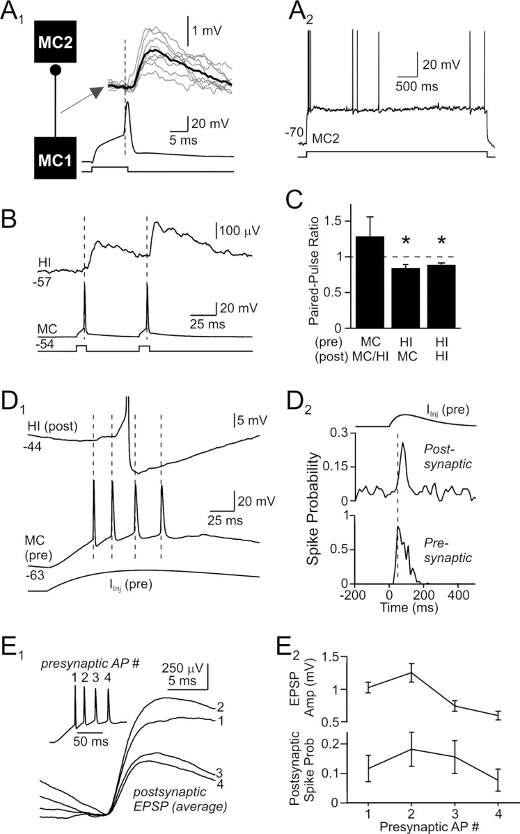
Functional effects of excitatory hilar synapses. A1, Presumptive mossy cell-to-mossy cell excitatory connection with a mean onset latency of 2.5 ms. Average postsynaptic response shown in bold trace superimposed on example unitary responses (gray traces). A2, The postsynaptic hilar neuron (distance metric, 1.57; MC2) in this paired recording responded to a depolarizing step with an initial burst followed by irregular spiking and small AHPs, characteristic of mossy cells. B, Averaged postsynaptic response to paired-pulse activation of a mossy cell. Hilar EPSPs showed modest paired-pulse facilitation. C, Summary of short-term plasticity in hilar synapses. Inhibitory responses segregated by phenotype of postsynaptic target; excitatory connections pooled. *p < 0.05. D1, Spike transmission through mossy cell synapses. The presynaptic mossy cell (bottom traces) fired repetitively at ∼50 Hz when activated by an α-function current waveform, evoking a series of summating EPSPs and a single AP (truncated) in the postsynaptic hilar interneuron (top trace). D2, Summary plots of the probability of firing in the presynaptic and postsynaptic cells, versus time, when the presynaptic neuron was driven by trains of α-function current waveforms as in D1. Repetitive mossy cell firing at 50 Hz reproducibly evoked firing in postsynaptic neurons. E1, Average spike-aligned responses to the first 4 APs in the α-function-triggered presynaptic bursts. Example presynaptic mossy cell burst shown in inset. The EPSP evoked by the second presynaptic AP was larger than the response to the first AP; subsequent EPSPs were smaller than the initial EPSP response. E2, Summary plots of EPSP amplitude and postsynaptic spike probability versus presynaptic spike number. Same stimulus protocol as D1 and E1.
