Figure 2.
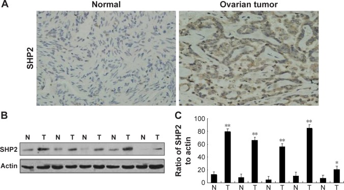
SHP2 overexpression in human ovarian tumor tissues.
Notes: (A) Representative images of immunohistochemical staining of human ovarian tissues (magnification, 400×). Immunohistochemical staining was performed to evaluate the SHP2 expression levels in 60 human ovarian cancer tissues (3+) and 60 matched normal ovarian tissues (1+). (B) Western blot analysis of SHP2 protein expression in human ovarian tissue specimens. (C) A semi-quantitative Western blot analysis of human ovarian tissue samples revealed that the levels of the SHP2 protein were significantly elevated in tumor tissues compared with normal tissues. Actin was used as an internal control. **P<0.01 compared with normal tissues, *P<0.05.
Abbreviations: N, normal; T, tumor.

