Figure 7.
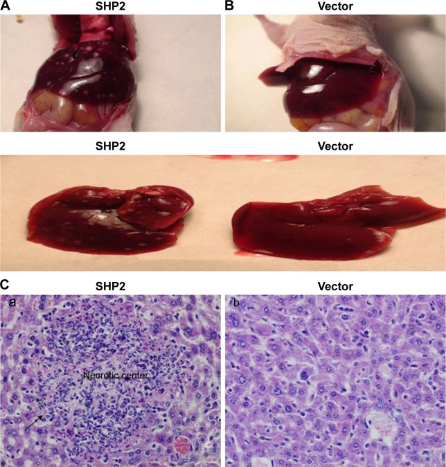
Effect of SHP2 overexpression on tumor metastasis.
Notes: A2780 cells were injected into nude mice. The mice were sacrificed 50 days later and tumor metastasis to distant organs was subsequently assessed. Anatomical images of liver metastasis in mice injected with the SHP2-overexpressing (A) or vector control (B) cells. Lower panel: metastases in the resected livers. (C) Cell morphology was evaluated using H&E staining; a:SHP2 group; b:Vector group. Liver tissues were fixed, sectioned, and stained with H&E, as described in the “Materials and Methods” section. A representative image of metastatic nodes in the livers of mice injected with SHP2-overexpressing cells is shown (magnification, 400×). A minimum of six regions from each tumor were examined.

