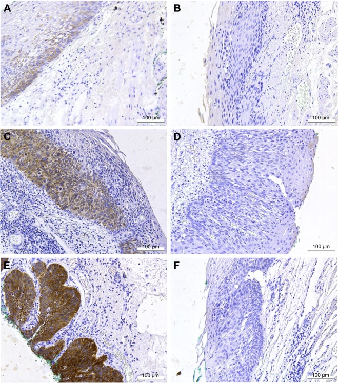
Figure 2.
Analysis of IGF2BP3 expression in HGIN by IHC.
Notes: All images were captured at 200× magnification. Positive (A) and negative (B) staining of IGF2BP3 in pT1a-EP (M1). Positive (C) and negative (D) staining of IGF2BP3 in pT1a-LPM (M2). Positive (E) and negative (F) staining of IGF2BP3 in pT1a-MM (M3). Positive staining of IGF2BP3 was characterized by a dark brown staining in the cytoplasm of the neoplastic cells.
Abbreviations: HGIN, high-grade intraepithelial neoplasia; IGF2BP3, insulin-like growth factor-II mRNA-binding protein-3; IHC, immunohistochemistry.

