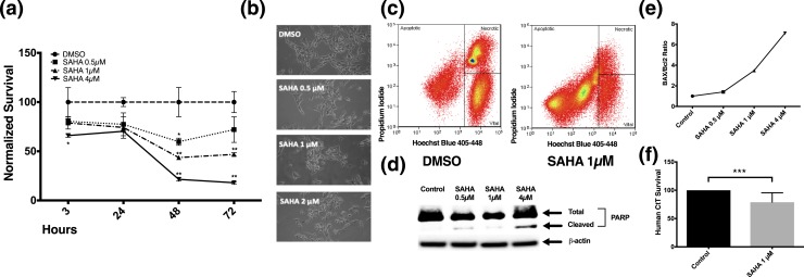
Figure 1.
SAHA reduced AtT-20 cell survival by apoptotic induction. Clinically achievable concentrations of SAHA (0.5 to 4 µM) significantly reduced the viability of transformed AtT-20 cells in concentration- and time-dependent manners following 3- to 72- hour treatment. (a) Time course of their survival, as assessed by MTT, was plotted. (b) Representative live microscopy images of AtT-20 cells exposed to various doses of SAHA for 24 hours. (c) FACS analysis revealed increased apoptosis in AtT-20 cells treated with SAHA (1 µM) for 24 hours. (d) Western blot analysis revealed an increase in cleaved PARP, a hallmark of apoptotic cell death, when AtT-20 cells were exposed for 24 hours to SAHA (0.5 to 4 µM). (e) SAHA (0.5 to 4 µM) resulted in an increase in the Bax/Bcl2 ratio at 24 hours in a dose-dependent manner in AtT-20 cells, suggesting that SAHA activates proapoptotic pathways. The Bax/Bcl2 ratio curve was generated from two independent experiments. (f) SAHA (1 µM) reduced cell survival at 24 hours in hCtT cells (n = 7). *P ≤ 0.05; **P ≤ 0.01; ***P ≤ 0.001 compared with corresponding control values. Horizontal bars represent mean ± standard deviation. DMSO, dimethyl sulfoxide.

