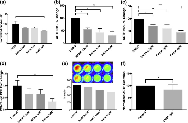
Figure 2.
Before apoptotic death, SAHA evoked robust and sustained declines in ACTH release and POMC transcription in AtT-20 cells. (a) Early (within 3 hours) exposure to SAHA (4 µM) significantly decreased the survival of AtT-20 cells, as observed in MTT assays. Percentage changes in ACTH secretion by AtT-20 cells upon exposure to increasing concentrations of SAHA for (b) 3 hours and (c) 24 hours; ACTH levels significantly decreased by clinically achievable concentrations of SAHA (0.5 to 4 µM) in both time groups (b, c). Each sample was run in duplicate in every experiment, and each experiment was repeated three times for the ELISA assay at 3 and 24 hours (b, c). (d) Physiologically available concentrations of SAHA led to transcriptional downregulation of POMC in AtT-20 cells after 24-hour exposure. (e) As inferred by the luciferase transfection study, the aforementioned effect was likely caused by suppression of the POMC promoter. (f) Expectedly, SAHA led to a significant decrease in ACTH levels after 24 hours of exposure on hCtT cells. *P ≤ 0.05; **P ≤ 0.01; ***P ≤ 0.001 compared with corresponding control values. Horizontal bars represent mean ± standard deviation.

