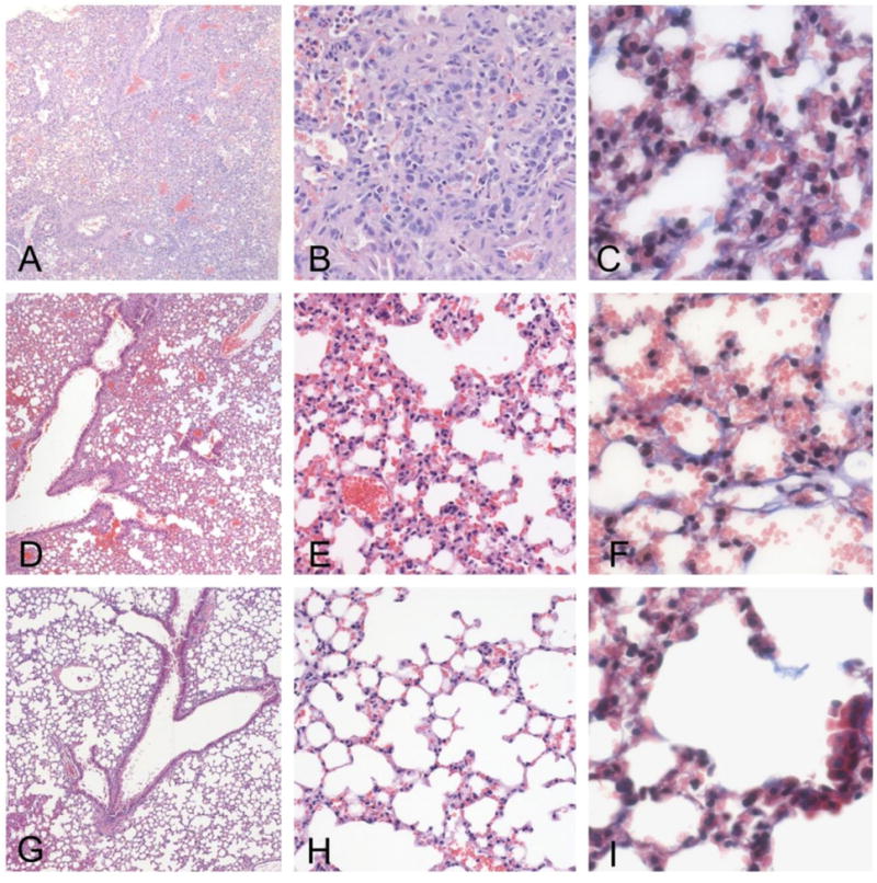
Figure 7. Histopathology of lung.

Lungs treated with bleomycin display severe injury, characterized by the obliteration of the alveolar space by proliferating stromal cells and a mixed inflammatory infiltrate, consisting primarily of macrophages. The infiltration was confluent and occupied sizable portions of the lung (a - X10, b - X20). Proliferation of fibrous tissue was observed along the alveolar walls by trichrome staining (c - 40X). Lungs of mice treated with bleomycin plus PGE2 and siRNA displayed a significant protective effect of treatment (7d - X10, e - X20). The alveolar structure was largely intact, with only small areas of infiltration and parenchymal consolidation. Only modest increase in connective tissue was observed by trichrome staining (d - X40). Lungs of control mice animals demonstrate a patent alveolar structure, with no inflammation, consolidation or fibrosis. (f - X10, g - X20, f - X40).
