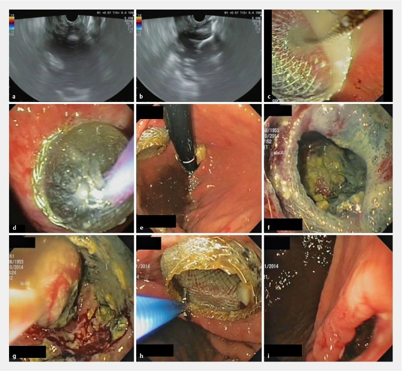
Fig. 3.

EUS-guided drainage with LAMS and necrosectomy in a patient with an infected WOPN. a EUS view of the WOPN, and b of the distal flange of the stent opened inside the collection. c Endoscopic view of the proximal flange of the stent. d Balloon dilation of the stent. e Endoscopic view of the stent correctly positioned with the proximal flange in the gastric cavity. f View through the stent of necrotic tissue. g Endoscopic view from inside the WOPN: retrieval of a fragment of necrosis with a standard net. h Stent removal with a biopsy forceps. i Cystogastrostomy immediately following stent removal.
