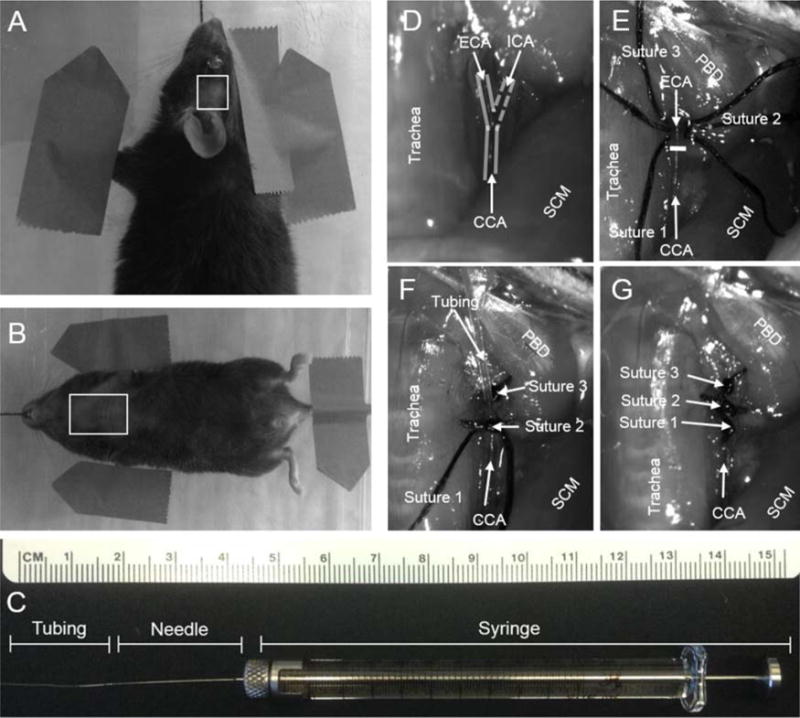
Fig. 1.

(A) Mouse in pronated position with box outlining shaved temporal region, temporal incision from lateral corner of left eye to medial region of left ear. (B) Mouse in supine position with box outlining shaved thoracic region. A midline incision is made from apex of ribcage to angle of mandible. (C) 100 μl syringe with 34 gauge and micro-angio tubing attached. (D) Outline of CCA/ECA/ICA with trachea and sternocleidomastoid (SCM) as landmarks. (E) Sutures 1–3 placement and white line marking CCA bifurcation, region framed by trachea, SCM and posterior belly of the digastric (PBD). (F) Micro-angio tubing inserted into ECA toward the CCA bifurcation and secured with suture 2. (G) ECA suture ligated.
