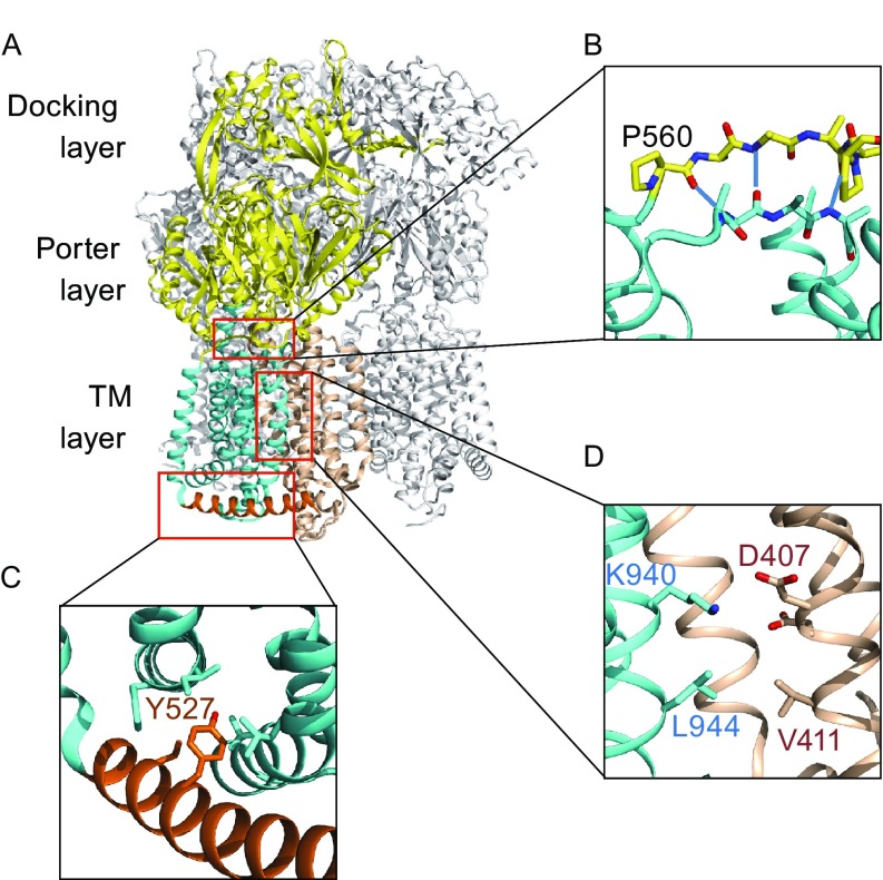
Figure 1.
AcrB structure and mutation sites. (A) Trimer crystal structure of E. coli AcrB (PDB ID: 4DX5). One protomer is shown in color, and the other two in grey. The NTM and CTM subdomains are shown in wheat color and cyan, respectively. The porter domain and docking domain are in yellow. The cytosolic-side amphipathic helix, α6-7, is colored in orange. (B) The signaling motif-C in CTM. The three main-chain H-bonds are depicted as dash-lines. Except proline residues, side-chains are omitted for clarity. (C) The α6-7 region. (D) Region of the titratable key residue(s)

