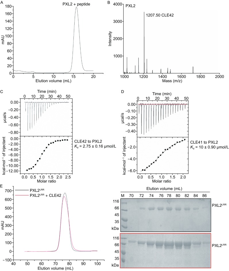
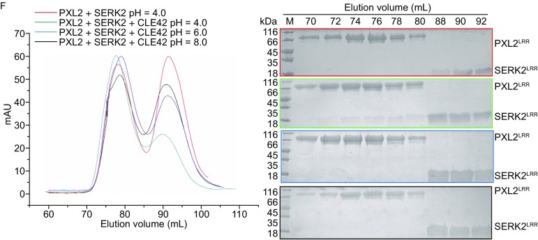
Figure 1.


CLE42 binding induces PXL2 LRR interaction with SERK2 LRR. (A) Gel-filtration chromatogram of the extracellular LRR domain protein of PXL2 (PXL2LRR) and a pool of synthesized peptides. The peak indicates the elution positions of PXL2LRR-peptide in gel filtration. The vertical and horizontal axes represent UV absorbance (280 nm) and elution volume (mL) respectively. (B) MALDI-TOF MS of the peak fraction of PXL2LRR-peptide shown in (A). The molecular weight of the peptide from the peak fraction (1207.50) indicated is equivalent to the theoretical weight of CLE42. The vertical and horizontal axes represent the intensity and molecular weight of MS respectively. (C) Measurement of the binding affinity between PXL2LRR and CLE42 by ITC. Top panel: twenty injections of CLE42 solution were titrated into PXL2LRR in the ITC cell. The area of each injection peak corresponds to the total heat released for that injection. Bottom panel: the binding isotherm for PXL2LRR-CLE42 interaction. The integrated heat is plotted against the molar ratio between CLE42 and PXL2LRR. Data fitting revealed a binding affinity of about 2.75 μmol/L. (D) Measurement of binding affinity between PXL2LRR and CLE41/TDIF by ITC. The assay was performed as described in (C). Data fitting revealed a binding affinity of about 10 μmol/L. (E) CLE42 binding induces no oligomerization of PXL2LRR. Left: gel filtration profiles of PXL2LRR in the presence and absence of CLE42. The vertical and horizontal axes represent ultraviolet absorbance (λ = 280 nm) and elution volume (mL), respectively. Right: Coomassie blue staining of the peak fractions of PXL2LRR shown in the left following SDS-PAGE. M, molecular weight ladder (kDa). (F) CLE42 induces PXL2LRR-SERK2LRR heterodimerization in solution at pH 4.0. Top panel, gel filtration profiles of PXL2LRR and SERK2LRR in the presence (slate at pH 4.0, blue at pH 6.0, black at pH 8.0), and absence (red) of CLE42. Right: Coomassie blue staining of the peak fractions of PXL2LRR and SERK2LRR shown in (F) following SDS–PAGE
