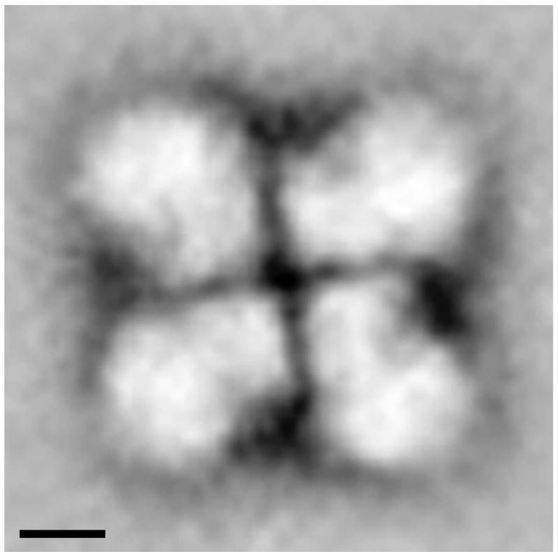
Fig. 12. The arrangement and orientation of native spliceosomes in the intact supraspliceosome.
Intact supraspliceosomes were classified by Correspondence Analysis and Hierarchical Ascendant Classification [80]. One of the classes is depicted showing the close contact between neighboring native spliceosomes in the center of the supraspliceosome, where the small subunits of the native spliceosomes are located. The contacts between the neighboring small subunits form a right-angled cross that reflects a four-fold arrangement. Scale bars represent 10 nm. Adapted from Ref [80].

