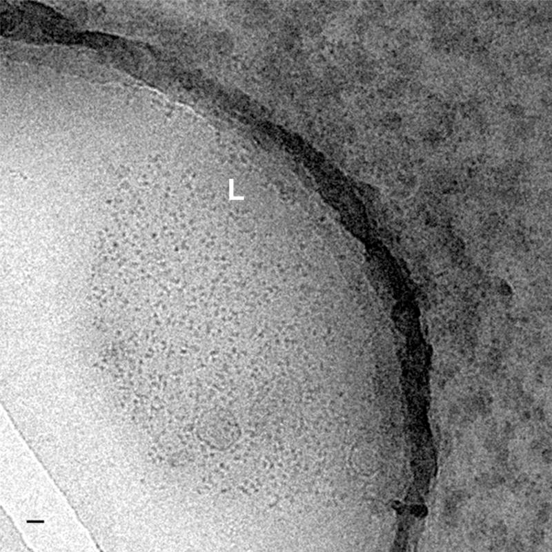
Fig. 3. Cryo-images of supraspliceosomes concentrated on a charged lipid monolayer.
A low magnification image of frozen-hydrated supraspliceosomes concentrated on a positively charged lipid monolayer (phosphatidylcholine and stearylamine) viewed inside a hole in the carbon-coated grid. The edges of the lipid film (L) are clearly visible and only very few particles are found outside the lipid layer. The bar represents 200 nm. Adapted from Ref [36].

