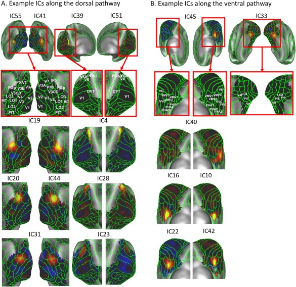
Figure 4.

Discrete ICs along the dorsal pathway and ventral pathway. A: The left panel shows the ICs that match well with existing lateral visual areas in the dorsal pathway. The right panel shows the ICs that match well with existing medial visual areas in the dorsal pathway. B: Example ICs that match well with existing medial visual areas in the ventral pathway such as VMV1–3, FFC, and VVC. The green lines mark the existing visual areal borders according to the MMP. For the sake of illustration, unilaterally distributed ICs (i.e., IC55 and IC41 at left and right MT, respectively) are shown together in one panel. [Color figure can be viewed at http://wileyonlinelibrary.com]
