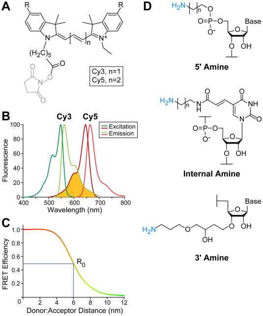
Figure 1.
Cy3 and Cy5 fluorophore characteristics and attachment to RNA for FRET studies. (A) Chemical structures of Cy dyes frequently used for FRET with NHS leaving groups (grey) for fluorophore attachment. R represents sulfonate groups typically added to Cy dyes to increase solubility. (B) Excitation (Ex) and emission (Em) spectra for Cy3 and Cy5 fluorophores. The spectral overlap between Cy3 emission and Cy5 excitation is shown (yellow). Spectra obtained from GE Healthcare Life Sciences. (C) Example plot of the FRET between a Cy3:Cy5 pair. At 0.5 FRET efficiency, the fluorophore pair distance is equal to the Förster distance, R0, which is ∼6 nm for Cy3:Cy5 [32]. (D) Examples of commercially available options for fluorophore labeling RNAs either at the 5′ or 3′ ends or internally. Chemical structures shown are those commercially available from Integrative DNA Technologies.

