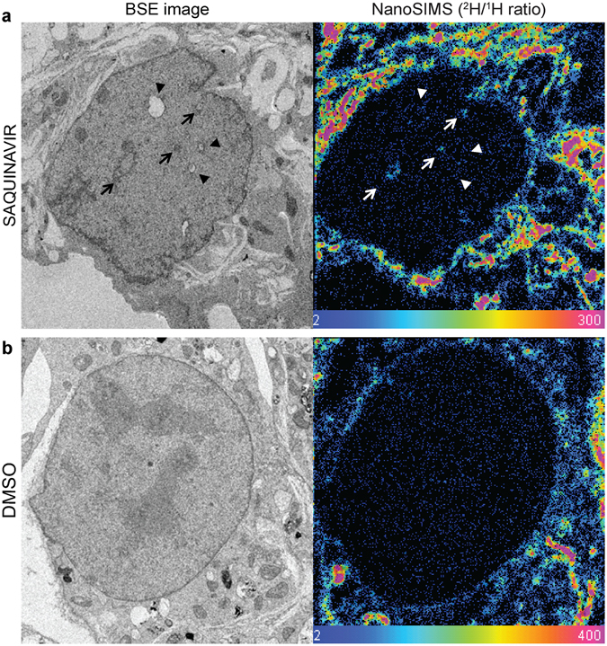
Figure 4.

Detection of nascent phospholipids during NR formation by pulse labelling with deuterated stearate. (a) Representative backscattered electron (BSE) image of a saquinavir-treated mouse preadipocyte with black arrows indicating NR tubules with a corresponding image from NanoSIMS showing enrichment of deuterium signal (2H/1H ratio) indicating nascent fatty acyl species, also at the NR foci (indicated with white arrows); black arrowheads in BSE image correspond to NR presumably present in the nucleus prior to 2H pulse, hence not showing enrichment in deuterium signal in the NanoSIMS image (white arroheads). (b) BSE and NanoSIMS images of a control cell treated with DMSO vehicle showing lack of NR tubules and no deuterium enrichment within the nucleus, and undistorted nuclear boundary. Colour scale of NanoSIMS images 2–400 equals 0.02–4% of 2H/1H.
