
Figure 5.
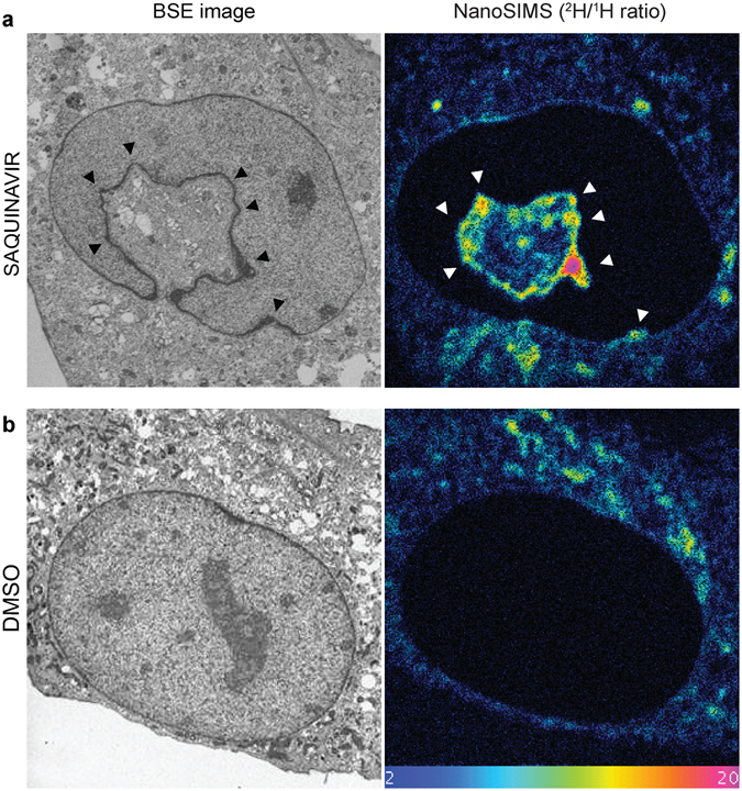
Detection of nascent phospholipids by pulse labelling with deuterated choline during NR formation. (a) Representative backscattered electron (BSE) image of a saquinavir-treated mouse preadipocyte (black arrowheads point towards high membrane curvature indicating NR initiation sites) with a corresponding image from NanoSIMS showing enrichment of deuterium signal (2H/1H ratio) revealing nascent phospholipids incorporated directly into forming NR (white arrowheads). (b) BSE and NanoSIMS images of a control cell treated with DMSO vehicle showing lack of NR tubules and no deuterium enrichment within the nucleus, and undistorted nuclear boundary. Colour scale of NanoSIMS images 2–20 equals 0.02–0.2% of 2H/1H ratio.