
Figure 6.
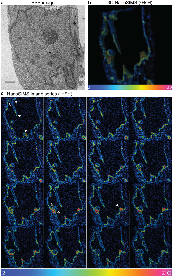
3D NanoSIMS of nascent phospholipid distribution during NR formation. (a) Representative backscattered electron (BSE) image of a saquinavir-treated mouse preadipocyte pulsed labelled with deuterated choline; scale bar 2 µm. (b) 3D reconstruction of NanoSIMS analysis of subsequent layers of the specimen shown in (a). (c) Panel of NanoSIMS images used for the 3D reconstruction shown in (b); examples of tip and base region of NR tubules are indicated by arrowheads and arrows, correspondingly; a single image corresponds to ~10 nm specimen thickness. Colour scale of NanoSIMS images 2–20 equals 0.02–0.2% of 2H/1H ratio. See corresponding Supplementary Movies 2 and 3.