
Figure 2.
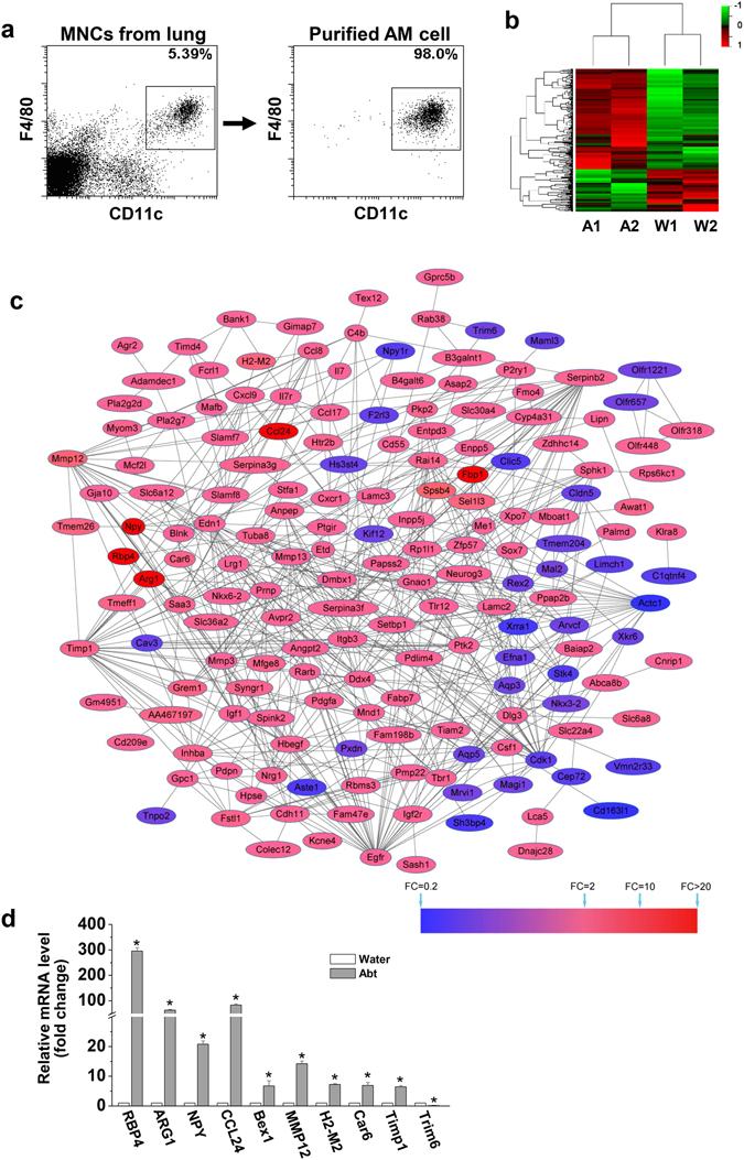
Altered gene expression in alveolar macrophages isolated from Abt mice. The mice in Abt group were given antibiotics for five weeks. Purified alveolar macrophages (F4/80hi CD11chi) were analyzed by GeneChip. (a) Alveolar macrophages (F4/80hi CD11chi) were sorted from lung MNCs. (b) The heatmap showed the differential expression of the indicated genes between the Abt group and the control group. DEGs were determined by Limma, and were row-based median normalized (2 samples/group, 15 mice/sample). (c) Protein-protein interaction (PPI) networks of the DEGs identified in the Abt group compared with the control. The red nodes indicate the up-regulated DEGs and the blue nodes indicate the down-regulated DEGs. Proteins associated with each other are linked by an edge. The degree of color represents the DEG fold change in reference to the scaleplate. (d) The mRNA expression levels of selected DEGs in the sorted macrophage cells (F4/80hi CD11chi) were measured by real-time PCR (n = 3). *p < 0.05 compared with the control group.