
Figure 4.
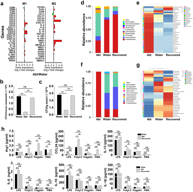
Recovered commensal bacteria restore the functions of alveolar macrophages in Abt mice. The mice in Abt group were given antibiotics for five weeks. In the recovered group, the mice were given antibiotics for five weeks and then co-housed with the wild type mice for four weeks. Purified alveolar macrophages (F4/80hi CD11chi) were analyzed. (a) M1 and M2 specific gene expression in alveolar macrophages from Abt mice was compared with that in control mice by GeneChip analysis. (b and c) Bacterial loads in the upper respiratory tract and stool from the recovered mice compared with those in the control mice (n = 3/group) were measured by BAP culture. (d and e) Relative abundances (d) and a clustering map (e) for the bacteria in the upper respiratory tract were determined by 16S rRNA analysis (3 samples/group, 10 mice/sample). (f and g) Relative abundances (f) and a clustering map (g) for the bacteria in the stool were determined by 16S rRNA analysis (3 mice/group). (h) Purified alveolar macrophages (F4/80hi CD11chi) were stimulated for 48 h in the indicated culture medium (1 µg/ml LPS, 100 µg/ml PolyI:C, 1 µg/ml flagellinor 20 ng/ml PMA) and then analyzed for the protein expression by ELISA (Arg1 and CCL24) and CBA (IL-13, IL-6, IL-10 and IL-1β) (n = 3). The data are shown as the mean ± SEM. *p < 0.05, **p < 0.01 compared with the control group.