Figure 5.

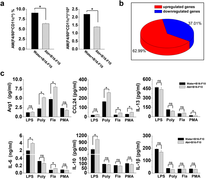
Decreased numbers but M2 polarization of alveolar macrophages in Abt mice challenged with B16/F10 melanoma. The mice were given antibiotics for five weeks and then challenged with B16/F10 cells (1 × 10⁵ cells/mouse, i.v.). On day 17 after the B16/F10 challenge, the lung MNCs were isolated. (a) Frequency and number of alveolar macrophages (F4/80hi CD11chi) in the lung MNCs from mice challenged with B16/F10 cells (n = 5). (b) Purified alveolar macrophages (F4/80hi CD11chi) were analyzed by GeneChip. The pie chart showed the distribution of DEGs in Abt group compared with the control. Unknown and duplicated genes were filtered for the analysis (2 samples/group, 15 mice/sample). (c) Purified alveolar macrophages (F4/80hi CD11chi) (3 × 10⁵ cells/well) were stimulated for 48 h in the DMEM containing 10% FBS with the stimulation of 1 µg/ml LPS, 100 µg/ml PolyI:C, 1 µg/ml flagellin or 20 ng/ml PMA, and then culture supernatants were analyzed for the cytokine expression by ELISA (Arg1 and CCL24) and CBA (IL-13, IL-6, IL-10 and IL-1β) (n = 3). The data are shown as the mean ± SEM. *p < 0.05 compared with the control group.
