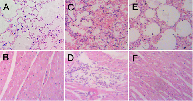
Figure 2.

Histopathologic appearance of tissues of the mink. (A) Lung tissue taken from a control mink on day 6 p.i. (B) Heart tissue taken from a control mink on day 6 p.i. (C) Lung tissue taken from a inoculated mink on day 6 p.i., characterized by large area of bleeding, thickening of the alveolar septa. (D) Heart tissue taken from a inoculated mink on day 6 p.i., characterized by infiltration of inflammatory cells. (E) Lung tissue taken from an exposure mink on day 6 p.i., characterized by slight thickening of the alveolar septa. (F) Heart tissue taken from an exposure mink on day 6 p.i., no obvious histologic lesions were found. HE stain. Original magnification was ×200 for all images.
