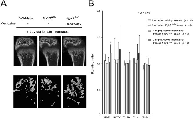
Figure 4.

Bone mineral density is enhanced in meclozine-treated Fgfr3 ach mice. (A) Representative reconstructed micro-CT images of the distal femur at the end of treatment revealed that metaphyseal bone mineral density was increased after meclozine treatment. Upper panels show 2D images of the distal femur and lower panels show 3D images of metaphyseal trabecular bone. (B) The average relative ratio of bone mineral density (BMD) was increased after meclozine treatment while there were no statistical differences in bone volume/total volume (BV/TV), trabecular thickness (Tb.Th), trabecular number (Tb.N), and trabecular separation (Tb.Sp). Relative values were calculated based on those of untreated Fgfr3 ach mice. Mean and SD are indicated. Statistical significance was analyzed by the unpaired t test for each dose of meclozine-treated Fgfr3 ach mice (n = 6 and 5 for 1 and 2 mg/kg/day of meclozine, respectively) or untreated wild-type mice (n = 10) versus untreated Fgfr3 ach mice (n = 9).
