
Figure 4.
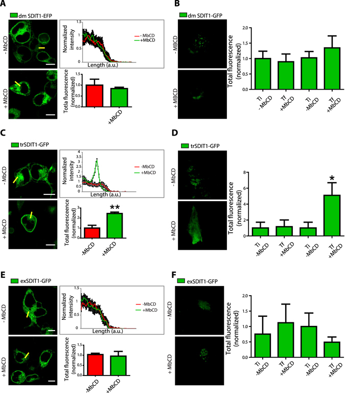
The extracellular CRAC domain from SIDT1 is part of a vesiculation signal required for translocation to the plasma membrane. (A) Representative confocal images of HEK293 cells expressing the SIDT1 double mutant from the CRAC domains (dmSIDT1). The right panel shows the line analysis illustrating the lack of translocation of the dmSIDT1 upon MβCD treatment. The lower panel shows the mean ± standard deviation of the fluorescence intensity in the line before and after MβCD treatment obtained from at least 40 independent experiments. (B) Total internal reflection fluorescence microscopy (TIRFM) representative images obtained with HEK293 cells expressing the dmSIDT1. The right panel shows the mean ± standard deviation from the total fluorescence of at least 30 independent measurements. (C) Representative confocal images of HEK293 cells expressing the SIDT1 transmembrane mutant (tmSIDT1). Notice the normal translocation to the plasma membrane after MβCD treatment. (D) Total internal reflection fluorescence microscopy (TIRFM) representative images obtained with the trSIDT1 mutant. (E) Representative confocal images of HEK293 cells expressing the exSIDT1 mutant. Notice that this mutant does not translocate to the plasma membrane after MβCD treatment. (F) Total internal reflection fluorescence microscopy (TIRFM) representative images obtained with the exSIDT1 mutant. Ti, the initial time of recording and Tf, final recording time. Total fluorescence normalized to Ti. In all panels scale bar: 10 microns.