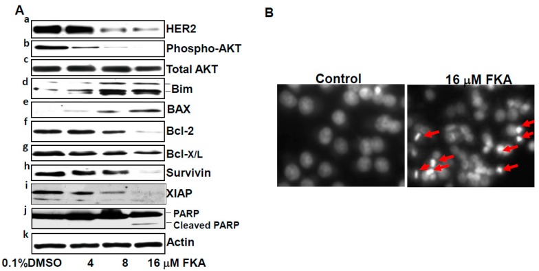
Figure 4.
FKA induces apoptosis in HER2-overexpressing SKBR3 breast cancer cell lines via downregulation of HER2, phosphorylated AKT, Bcl2, Bcl-X/L, survivin, and XIAP and upregulation of Bim and BAX. (A) SKBR3 cells were treated with indicated concentrations of FKA for 24 h. Western blotting analysis of HER2, phosphorylated AKT, AKT, Bim, BAX, Bcl-2, Bcl-X/L, survivin, XIAP, and PARP is shown by a representative blot from three experiments. β-actin was detected as a loading control; and (B) DAPI staining of nuclear morphology under fluorescence microscope (magnification: 200×). Arrows indicate cells with nuclear condensation and fragmentation, which were counted as apoptotic cells.

