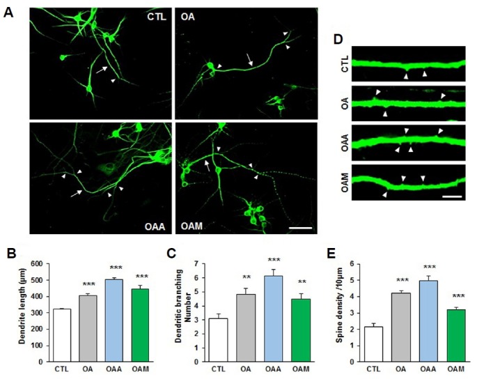
Fig. 2. OA, OAA and OAM increase neurite length and spine density.

(A) Hippocampal primary neurons were exposed to OA, OAA or OAM for 72 h. The neurite length (arrow) and number of branches (arrowheads) were counted in neurons immunostained for MAP2. Images were captured at a magnification of ×60, using a confocal microscope. Scale bar, 50 μm. (B, C) Treatment with OA, OAA and OAM significantly increased neurite length and number of neurite branches (n = 10–16 neurons/condition, two independent cultures). (D) Representative images are shown of high-magnification Z-stack projections of neurite spine density. Arrowheads indicate the density of neurite spines. Scale bar, 5 μm. (E) The density of neurite spines was significantly increased by treatment with OA, OAA and OAM (n = 15–22 neurons/condition, two independent cultures). Results are mean ± SEM. Student’s t-test *p < 0.05, **p < 0.01, ***p < 0.001 compared to control (CTL).
