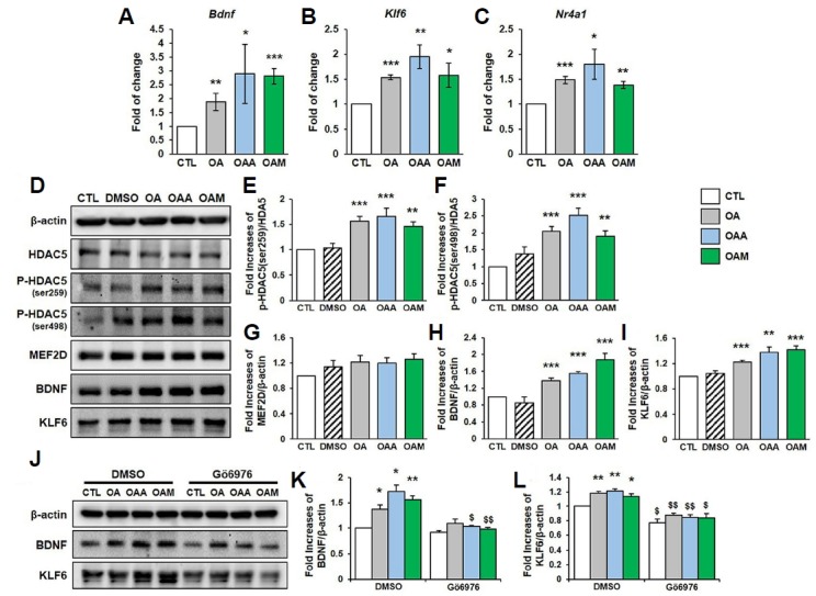
Fig. 5. OA, OAA and OAM induce MEF2 target gene expression via HDAC5 dissociation in rat hippocampal neurons.

(A–C) The expression of Bdnf, Klf6 and Nr4a1 mRNA levels in hippocampal neurons after treatment with OA, OAA or OAM for 72 h. Results of quantitative PCR were normalized to the level of β-actin and are shown as fold changes relative to control neurons (n = 3 independent cultures). (D–I) Hippocampal neurons were treated with OA, OAA or OAM for 72 h. HDAC5 phosphorylation, and expression of MEF2D, BDNF and KLF6 proteins were analyzed by Western blot (n = 4 independent experiments). (J–L) Hippocampal neurons were pretreated with DMSO or Gö6976 (1 μM) for 30 min, and then exposed to OA, OAA or OAM for 72 h. The expression of BDNF, KLF6 and β-actin in whole cellular extracts were determined by Western blot analysis (n = 3 independent experiments). Results are mean ± SEM. Student’s t-test, *p < 0.05, **p < 0.01, ***p < 0.001 compared to control (CTL). $p < 0.05, $$p < 0.01, $$$p < 0.001 compared to of the group that received DMSO treatment (DMSO).
