
Fig. 3. Activation of 5-HT6Rs increases the number and size of dendritic protrusions in cultured hippocampal neurons.
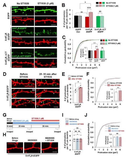
(A–F) Expression of 5-HT6R-EGFP, but not 5-HT6R-ΔCT-EGFP, exhibits ST1936-mediated morphological changes of dendritic protrusions. (A) Confocal images of neurons expressing EGFP, 5-HT6R-EGFP or 5-HT6R-ΔCT-EGFP together with mCherry. Neurons were transfected at DIV10–13 and imaged at DIV12–15. (B) Dendritic protrusion density was analyzed. Around 1,800 to 4,500 μm dendritic length for each was analyzed from 12, 12, 18, 16, 25, 23 neurons from left to right. (C) Cumulative distribution of individual dendritic protrusion size. Insets display means ± S.E. of individual dendritic protrusions. n = 1022, 746, 1391, 1554, 2239, 2038 protrusions from 12, 12, 18, 16, 25, 23 neurons from left to right. (D) Confocal live–cell images before and 25–30 minutes after ST1936 treatment. Three pairs of representative images (D1, D2, and D3) were shown. Neurons were co-transfected with 5-HT6R-EGFP and mCherry at DIV13 and live–cell imaged at DIV15. White and green dots indicate newly formed and growing dendritic protrusions, respectively, after ST1936 treatment. (E) Dendritic protrusion density was analyzed. Around 700 μm dendritic length was analyzed from 3 neurons. (F) Cumulative distribution of individual dendritic protrusion size. n = 446, 594 protrusions from 3 neurons. Student’s paired t-test was used for (F). (G–J) Blockade of 5-HT6Rs inhibits ST1936-mediated morphological changes of dendritic protrusions in cultured hippocampal neurons. (G) Experimental schematic diagram for effect of 5-HT6R inhibitor on ST1936-mediated morphological changes of dendritic protrusions. (H) Representative confocal images from live-cell imaging experiment from (G). (I) Dendritic protrusion density was analyzed. Around 600 μm dendritic length was analyzed from 4 neurons. (J) Cumulative distribution of individual dendritic protrusion size. n = 290, 291, 287 protrusions from 4 neurons. Student’s paired t test was used for (I). NS: not significant, *P < 0.05, **P < 0.01, *** P < 0.001. Scale bar, 5 μm.