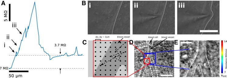
Fig. 2.
Lateral navigation around obstructions prevents persistent pipette resistance increase caused by penetration of blood vessels in vitro. A: resistance trace as a function of distance as a pipette pierces a blood vessel under high positive pressure. A residual resistance increase of 3.7 MΩ remains after the vessel is punctured. B: IR DIC images showing the pipette encountering and deforming the blood vessel. Scale bar, 50 µm. C: schematic of scanning ion conductance microscopy (SICM) mapping of a blood vessel proceeds from a central point. Samples are collected randomly from a grid area 20 × 20 μm at 2-μm resolution. D: the entire blood vessel and surrounding milieu are shown under IR DIC. Scale bar, 20 μm. E: resistances mapped as a function of grid position, clearly showing increased resistance when the pipette is above the blood vessel. Scale bar, 10 μm; 2× interpolation.

