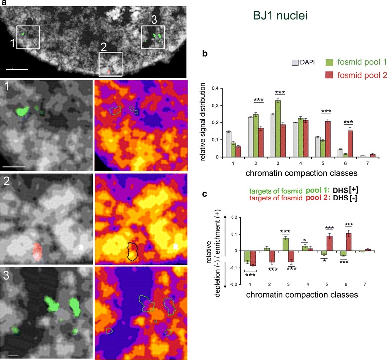
Fig. 4.

3D nuclear topography and quantitative mapping of ~40-kb targets of DHS[+] and DHS[−] regions in BJ1 nuclei. a Part of a SIM light-optical section from a whole nucleus acquisition with framed areas indicating representative inset magnifications 1–3. DAPI-stained DNA after intensity classification shown as gray gradations and color heat maps, respectively. Segmented signals delineating targets of fosmid pool 1 (DHS[+], green) and fosmid pool 2 (DHS[−], red) show a preferential localization of pool 1 signals DHS[+] in zones of low DAPI intensity and of pool 2 (DHS[−]) within the more compacted core of CDCs as shown by outlined signals in color heat maps: pool 1 = green, pool 2 = black). Scale bar 2 µm, insets 0.5 µm. b Quantified distributions (N = 25 nuclei) of fosmid pool 1 (DHS[+], green) and pool 2 (DHS[−], red) within respective chromatin compaction classes (all classes shown in gray) confirm the significantly distinct topography for DHS[+] and DHS[−] associated signals with a shift of DHS[−] sites toward higher compaction classes. c Quantified levels of relative enrichment (positive values) or depletion (negative values) of fosmid pool 1 and pool 2 signals within chromatin compaction classes. Error bars = standard deviation of the mean.*p ≤ 0.05; **p ≤ 0.01; ***p ≤ 0.001
