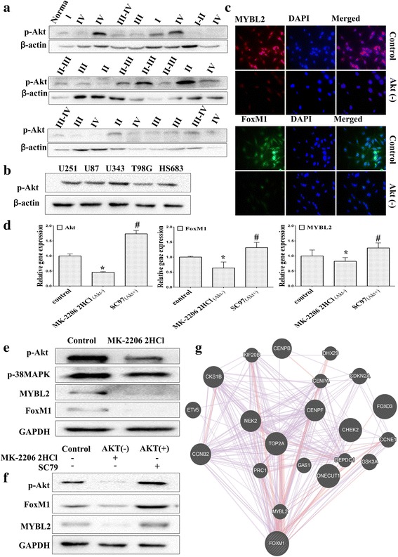
Fig. 8.

MYBL2 and FoxM1 are activated by Akt signaling pathway. a The baseline expression of p-AKT was determined by Western blotting in 26 glioma specimens and 1 normal tissue. b The expression of p-Akt was determined in glioma cell lines using Western blotting analysis. c-e U251 cells were treated with PAMK-2206-2HCL for 24 h. The expression of FoxM1 and MYBL2 were detected by immunofluorescence (c) real-time PCR (d) and Western blotting (e). f U251 cells were treated with PAMK-2206-2HCl or SC79 for 24 h. The expression of FoxM1 and MYBL2 were detected by western blotting. g The molecular functional network map of canonical pathways including coexpression, physical interaction, and predicted networks of FoxM1 analyzed by GeneMANIA (http://genemania.org/) tool.*P < 0.05 represent MYBL2 group vs. NC group; #P < 0.05 represent FoxM1 group vs.NC group
