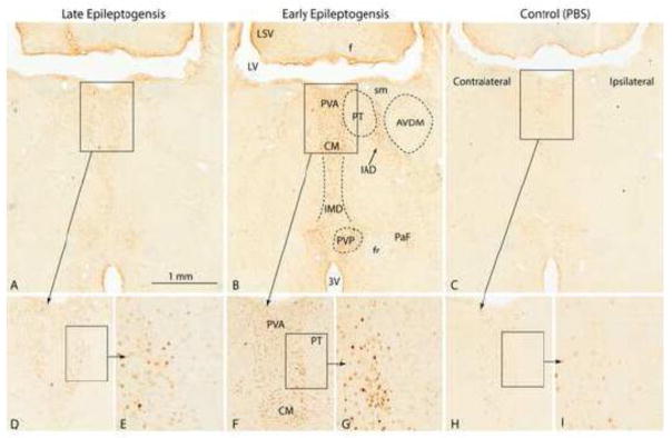
Figure 5.
C-Fos staining in midline thalamic nuclei of representative MSO-infused rats during early [B, F, G; Rat D (m)] and late epileptogenesis [A, D, E; Rat O (m)]. A stained nonepileptic, PBS-infused control [Rat C (pbs)] is shown in C, H and I. There is intense staining of neurons in the anterior part of the periventricular thalamic nucleus (PVA), the central medial thalamic nucleus (CM), the intermediodorsal thalamic nucleus (IMD), the interanteromdial thalamic nucleus and the posterior part of the paraventricular thalamic nucleus (PVP) during early epileptogenesis and slightly less intense staining in the same areas during late epileptogenesis. Weak staining of very few cells were occasionally encountered in the PBS control (C, H, I). Abbreviations: 3V, third ventricle; AVDM, anteroventral thalamic nucleus, dorsomedial part; f, fornix; fr, fasciculus retroflexus; LSV, lateral septal nucleus, ventral part; LV, lateral ventricle; PT, parathenial thalamic nucleus; sm, stria medullaris.

