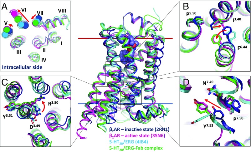
Fig. 3.
Receptor activation-related features of the 5-HT2B/ERG-Fab complex. Superposition of β2AR-Gs active state (magenta; PDB ID code 3SN6), β2AR inactive state (dark blue; PDB ID code 2RH1), 5-HT2B/ERG (light blue; PDB ID code 4IB4), and 5-HT2B/ERG-Fab complex (green). (A) View from the intracellular side. (B) PIF motif. (C) D(E)RY motif. (D) NPxxY motif. Major activation-related rearrangements observed in β2AR are shown as red arrows.

