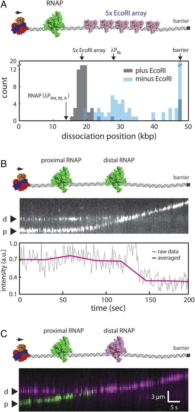
Fig. 5.
RecBCD sequentially disrupts tandem RNAPs. (A) Experimental schematic (Upper) and resulting dissociation position distribution data (Lower) for Qdot-tagged RNAP pushed into 5× EcoRI arrays by RecBCD. (B) Representative one-color kymograph showing two Qdot-tagged RNAP complexes (promoter-bound holoenzyme) being pushed into one another by unlabeled RecBCD and the corresponding graph showing the cumulative fluorescence intensity of the Qdot-tagged proteins. (C) Representative two-color kymograph showing two Qdot-tagged RNAP complexes (promoter-bound holoenzyme) being pushed into one another by unlabeled RecBCD; similar outcomes were observed in most (n = 50/52) two-color RNAP collisions, and in the remaining cases, RecBCD pushed both proteins (n = 2/52). p and d represent proximal and distal RNAP, respectively. In B and C, reactions were initiated by the addition of 1 mM ATP into the RecBCD buffer (40 mM Tris⋅HCl, pH 7.5, 2 mM MgCl2, 0.2 mg mL–1 Pluronic), and gaps in the RNAP trajectories result from Qdot blinking.

