Fig. 5.
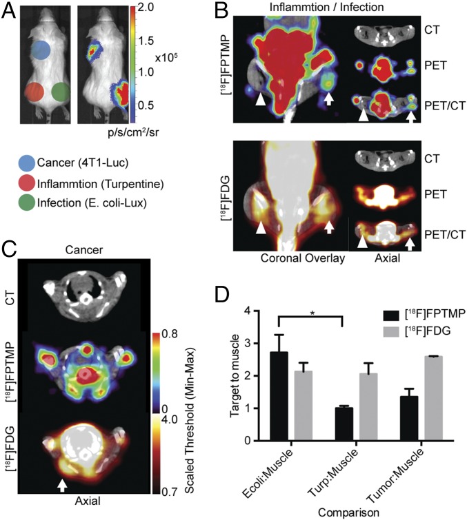
[18F]FPTMP detects live bacterial infection, not cancer or inflammation. (A) Schematic of locations of infection, inflammation, and cancer in a BALB/c mouse. Mouse mammary carcinoma cells (4T1Luc+, 1 million) were injected s.c. over the left shoulder of the mouse. Chemical inflammation with turpentine (30 μL IM) was induced 2 d before [18F]FPTMP imaging and 3 d before FDG imaging. Live E. coli bacteria (1 × E8 cfu i.m.) were injected into the right lower leg. (Right) Bioluminescent imaging of a representative animal after D-luciferin injection (1 mg, i.p.) illustrating active bacterial infection in the right leg and live tumor cells in the left shoulder region. (B) A representative animal after [18F]FPTMP, ∼200 μCi i.v., shows uptake in the infected hindlimb muscle (arrow) 4 h after infection, but not in the area of turpentine injection (arrowhead). Next-day imaging with [18F]FDG, ∼300 μCi i.v., shows uptake in both infection and chemical inflammation 1 h after injection. (C) The same animal as in B is shown after [18F]FPTMP and [18F]FDG injection at the site of a 4T1 tumor. There is increased [18F]FDG uptake at the site of the tumor and no specific signal from [18F]FPTMP. (D) Quantification of the data from B and C. Error bars represent the SD (n = 4). *P < 0.05.

